Perfusion is a window into tissue function
Start using our software to process your DCE-MRI and DSC-MRI data accurately and precisely. Test out the state-of-the-art algorithms.
Get startedFeatures
Flexible input format
Clinical and preclinical DICOM data from various scanner vendors, Bruker raw-data format, NIfTI format, and custom formats on request. DCE-MRI, DSC-MRI or simultaneous DCE-DSC-MRI.
Tailored AIF estimation
Pick the AIF that best suits your dataset: choose from our predefined set of AIFs, measure and scale the AIF within an artery, or use our blind-deconvolution estimation technique.
Automation of processing
It's up to you whether you want to process the data automatically or take control of the processing steps.
Regularized parameter estimation
We offer a unique algorithm calculating spatially regularized perfusion-parameter maps to ensure more reliable and spatially coherent results.
Statistical evaluation
To facilitate the analysis of your results, we provide basic statistical evaluation of perfusion maps along with exportable results tables.
Multiple pharmacokinetic models
The software includes all commonly used pharmacokinetic models - Patlak, Tofts, extended Tofts, 2CX, ATH, DCATH, GCTT.
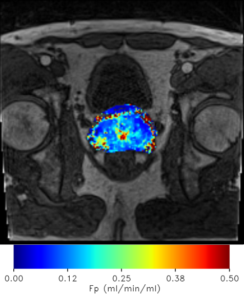
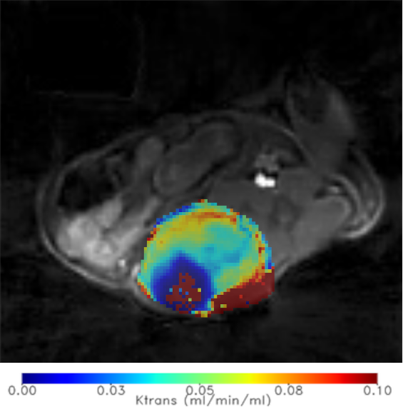
Clinical data
Diagnosis of prostate cancer using DCE-MRI.
Data from Masaryk Memorial Cancer Institute.
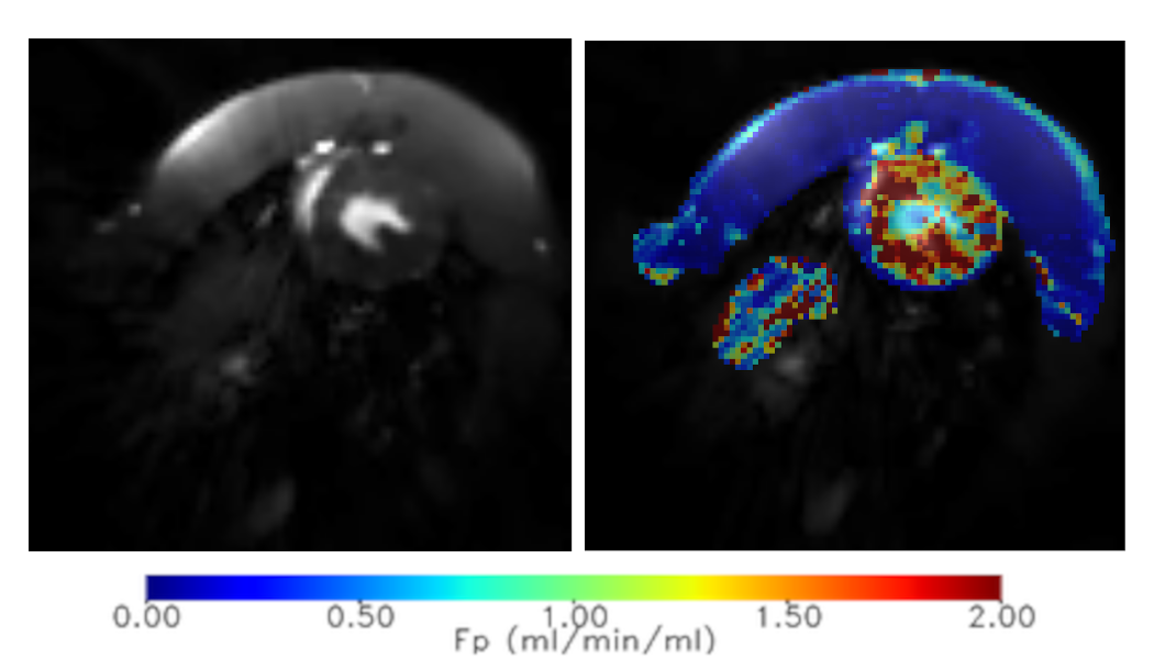
Preclinical data
Small-animal studies focusing on tumor-treatment develompment.
Synthetic data
Testing of new methods using custom simulated data.

Myocardial perfusion
Evaluation of myocardial perfusion using DCE-MRI.
Examples
Conversion to concentration
Checking the results
How to get started?
Contact us! We will create an account for you.
Contact Information
perflab@isibrno.cz
ISI of the CAS
Brno, Czechia